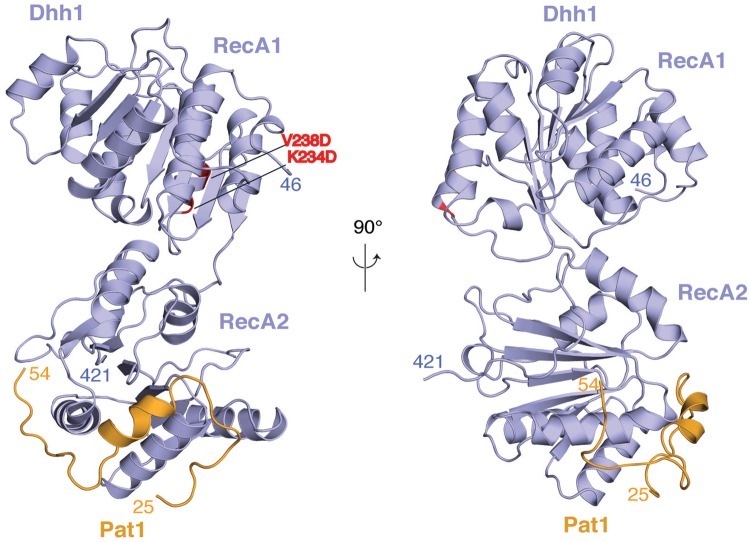
Figure 2.
Structure of the yeast Dhh1–Pat1 core complex. The two RecA domains of Dhh1 are in blue. The Dhh1-binding domain of Pat1 spans residues 24–54 (in orange). The structure is shown in two views related by a 90° rotation around a vertical axis. The N- and C-terminal residues ordered in the structure are indicated. The two residues of RecA1 mutated for crystallization (K234D and V238D, shown in red) are far from the RecA2 domain, where Pat1 binds.

