Fig. 4.

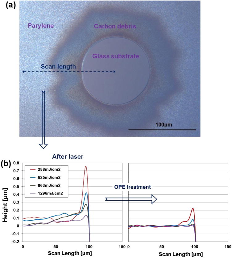
(a) Optical image of a laser-ablated spot (diameter of 100 μm) on a soda-lime glass. (b) Surface profiles of the edges of circles ablated using different laser fluences (left) and after 2 min of OPE treatment (right).

(a) Optical image of a laser-ablated spot (diameter of 100 μm) on a soda-lime glass. (b) Surface profiles of the edges of circles ablated using different laser fluences (left) and after 2 min of OPE treatment (right).