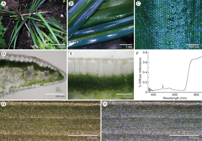
Fig. 1.
General appearance of M. caudata leaf structure by light microscopy, and optical properties. (A) Young plants growing in shade. (B) Intense blue–green iridescent colour from edge of leaf to midrib. (C) Reflected light on the surface of the leaf reveals iridescence of individual epidermal cells. (D) Transverse section of freshly sectioned leaf showing dense chloroplast concentration in palisade mesophyll, and rhomboidal epidermal cells. (E) Transverse section of epidermal cells shows thickened adaxial cell wall. (F) Diffuse spectral reflectance of typical iridescent blue leaf. Arrow indicates blue peak. (G) Leaf surface reflectance observed with a right circularly polarizing (RCP) filter. (H) Leaf surface reflectance observed with corresponding LCP filter.

