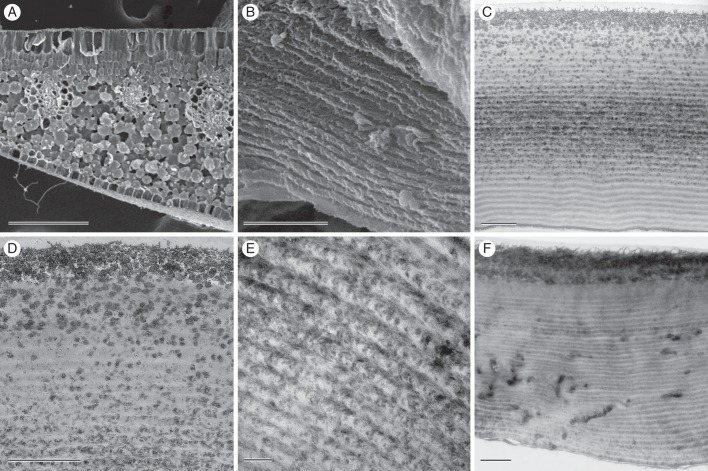
Fig. 2.
Electron microscopy of leaves of M. caudata. (A) SEM of leaf transverse section showing rhomboidal epidermal cells and densely packed palisade mesophyll cells. (B) Adaxial epidermal cell in SEM, revealing wall layering present through most of the width of the wall. (C) TEM of transverse adaxial epidermal cell wall shows layers of alternating electron translucency and a thicker, single granular layer near the surface. (D) Higher magnification TEM image near adaxial surface showing relationship of granules and helicoid layering. (E) TEM of adaxial wall near the middle of the adaxial wall, revealing helicoid appearance in finer detail. (F) TEM of adaxial epidermal cell wall in a non-iridescent portion of the leaf. Scale bars: (A) = 200 μm; (B–F) = 1 μm.

