Figure 4.

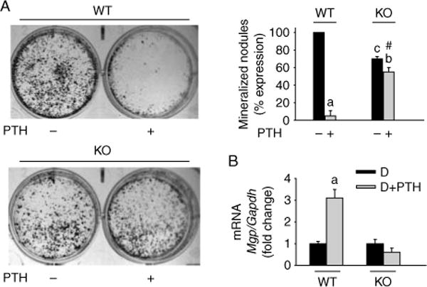
Effects of Mkp1 deletion on osteoblast mineralization and MGP mRNA expression with or without PTH treatment. Cells were isolated from calvaria of 11-week-old wild-type (WT) and Mkp1 knockout (KO) mice. The cells were differentiated with ascorbic acid and β-glycerophosphate for 21 days with or without 100 nM PTH. Mineralized nodule formation was examined by von Kossa staining as described in the Materials and Methods section. (A) Representative wells of 5–7 individual experiments with similar results for nodule formation are shown. The number of nodules was counted and plotted as percentage (%) expression of nodules with respect to PTH untreated WT cells from 5 to 6 individual experiments. Values are expressed as mean±S.D. a, P<0 001 versus untreated cells; b, P<0 01 versus untreated cells; c, P<0 01 versus untreated WT control cells; # P<0 001 versus PTH-treated WT control cells. The number of mineralized nodules in osteoblasts derived from KO animals without PTH treatment was 30% lower compared with PTH untreated WT cultures. (B) Mgp expression in primary calvarial osteoblasts derived from WT and Mkp1 KO mice at day 15 with and without PTH treatment. Total RNA was isolated from triplicate independent cultures. Results are graphically represented after normalization with Gapdh as fold change (mean±S.D.) with respect to PTH untreated differentiated cells from WT cultures. D, differentiated cells; a, P<0 001 versus D.
