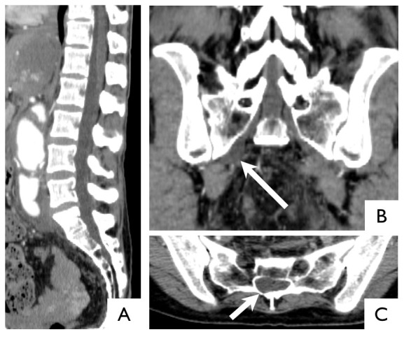
Figure 2. CT images of 46-year-old female with Loeys-Dietz syndrome.

Sagittal image of the normal dura (A). Coronal image of right lateral meningocele (arrow) (B). Axial image at S1 shows asymmetric dilatation of the dura (arrow) (C). In this case, visual inspection could detect dural ectasia, but quantitative evaluation could not.
