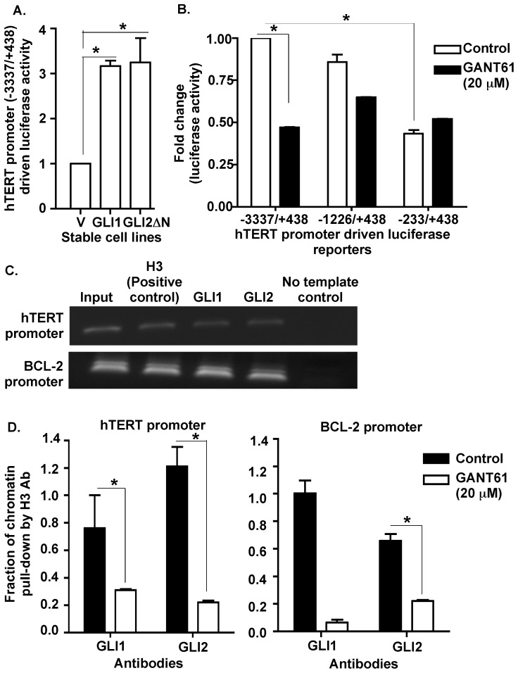
Figure 4. GLI1 and GLI2 proteins regulate hTERT promoter activity.
A: Stable derivatives of HT29 cells (described in Fig. 2C) were co-transfected with a full-length hTERT promoter driven luciferase (−3337/+438) reporter and renilla luciferase (pRL-TK) constructs for 24 hr. Lysates were prepared, and luciferase activity determined as described in Materials and Methods. hTERT promoter luciferase activity was normalized against renilla luciferase activity and is presented as mean ± SD, n = 3. B: HT29 cells were co-transfected with either the full length (−3337/+438) or upstream deleted mutants (−1226/+438 or −233/+438) of hTERT prom-luc reporters and pRL-TK followed by exposure to GANT61 (20 µM, 24 hr) and determination of luciferase activity. hTERT promoter luciferase activity was normalized against renilla luciferase activity and is presented as mean ± SD, n = 3. C: HT29 cells were employed for ChIP analysis using antibodies specific for GLI1, GLI2, or histone H3 (positive control, used for normalization). D: HT29 cells treated with GANT61 (20 µM, 24 hr) were similarly evaluated by ChIP analysis. Subsequent Real-Time PCR used primers that flanked the promoter regions of hTERT or the GLI target gene, BCL-2 (positive control). *p<0.05.

