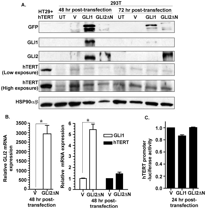
Figure 5. Over-expression of GLI1 or GLI2 in non-malignant 293T cells does not influence hTERT expression.
A: 293T cells were untreated (UT), transiently transfected with empty vector (V) or GFP-tagged GLI1 (GLI1) or GFP-tagged GLI2ΔN (GLI2ΔN). Expression of GLI1, GLI2 and hTERT was determined for 48 and 72 hrs by Western analysis. HSP90α/β was used as loading control and GFP was used to mark exogenously expressed GFP-tagged GLI proteins. Total cell lysate from HT29 cells stably expressing hTERT (HT29+hTERT) served as positive control. B: 293T cells transiently transfected with vector or GLI2 (as described in Figure 5A) were analyzed for GLI1, GLI2 and hTERT mRNA expression via qPCR. Data represent the mean ± SD of 3 determinations. C: 293T cells transiently co-transfected with FL-hTERT prom-luc, renilla luceferase reporters and vector, GLI1 or GLI2 (as described in Figure 5A). 24 hr post-transfection the cells were analyzed for luciferase activity. hTERT promoter-driven luciferase activity was normalized against renilla luciferase activity and is represented as mean ± SD, n = 3. *p<0.05.

