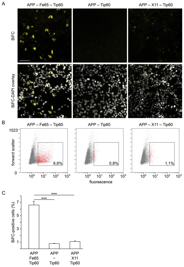
Figure 3. AFT-BiFC requires the presence of Fe65.
(A) After cotransfection of APP-YC, Tip 60-YN and HA-Fe65 the BiFC signal can be detected in a subset of cells in the confocal microscope. In contrast, cotransfection of APP-YC and Tip 60-YN alone does not lead to fluorescence complementation, which indicates absence of direct interaction between AICD and Tip60. Transfection of APP-YC and Tip 60-YN together with the AICD-binding protein MINT1/X11 that traps AICD in the cytosol also does not generate a BiFC signal. Lower panels show BiFC overlay with DAPI nuclear staining. Length of bar: 60 µm. (B) Representative BiFC-FC scatter plots of individual samples. Percentages refer to gated cells. Fluorescence intensity and forward scatter are depicted in arbitrary units. (C) BiFC-FC quantification of HEK293 cells cotransfected with APP-YC and Tip 60-YN together with or without HA-Fe65 or with MINT1/X11 (n=6 vs. 5 vs. 6, error bars represent SEM, *** p<0.005, U-test). The approximately 1% BiFC-positive cells in the APP-Tip60 and APP-X11-Tip60 conditions are most likely due to background autofluorescence of HEK cells as well as fluorescence complementation mediated by endogenous Fe65.

