
Figure 1. Flow cytometry-based detection of HC-04 cells infected with P. berghei ANKA GFP.
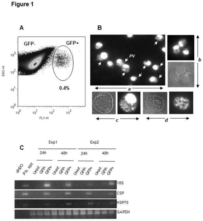
(A) HC-04 cells infected with P. berghei ANKA GFP sporozoites were detected by flow cytometry based on GFP expression. (B) GFP+ cells were isolated at 24 hrs post-infection by flow cytometry-based cell sorting and stained with DAPI (a, b). Parasitophorous vacuoles (PVs) are indicated by arrows. (c) GFP+ cells isolated at 48 hrs post-infection and stained with DAPI contain large PVs and some cells (d) resemble merosome-like structures. (C) mRNA expression of P. berghei ANKA GFP genes 18S, CSP and HSP70 was analyzed by RT-PCR in GFP+ and GFP- cells purified by sorting from infected cultures. Samples were collected at 24 and 48 hrs post-infection. HC-04 cells from non-infected cultures were used as a negative control. Data from two independent experiments are shown in the figure.