
Figure 2. MHC class I pathway gene expression in hepatocytes infected with P. berghei ANKA GFP.
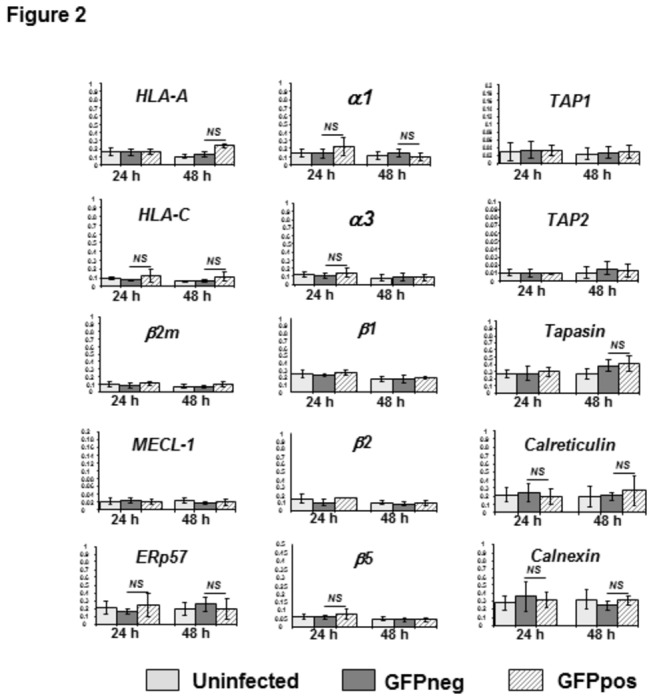
GFP+ and GFP- HC-04 cells were isolated by FACS sorting at 24 and 48 hrs post-infection from the same parasite-infected cell cultures. Uninfected cells were also subjected to sorting prior to mRNA isolation. Real time PCR analysis of mRNA expression was done for the 15 genes indicated. Each assay was performed in triplicate. Data are shown as expression units relative to GAPDH expression levels arbitrarily taken as 1 and represent the mean ± SD from 4 to 9 independent infection experiments and cell sorting procedures. NS, not significant.