
Figure 4. Circumsporozoite protein does not affect basal or inducible expression of MHC class I.
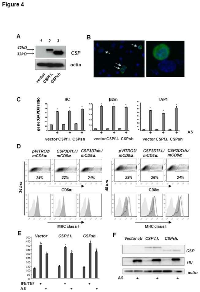
(A) Expression of the full length (CSPf.l.) or mature “short” (CSPsh) form of the P. falciparum 3D7 CSP protein was detected by western blotting 24 hrs after transfection of HC-04 cells. (B) Cellular distribution of full length CSP 24 hrs post-transfection was visualized with a CSP-specific antibody (green) and immunofluorescence microscopy. Nuclei were stained with DAPI (blue). Arrows indicate transfected cells. (C) Real-time PCR analysis of MHC class I heavy chain, β2-microglobulin and TAP1 gene expression in cells transiently expressing of P. falciparum CSP. HC-04 cells transfected with the control vector plasmid or plasmid encoding either full length or short CSP were treated with AS (10% v/v) 4 hrs post transfection or left untreated. Transfected cells were isolated 24 hrs later using surface expression of mouse CD8α as a marker. Mean ± SD of the assay triplicates. All p* <0.0002 and indicated differences between control and AS-treated samples. (D) Percentages of cells transiently expressing CSP were identified by CD8α co-expression (upper panels) and MHC class I was assessed by flow cytometry (lower panel, light gray histograms - isotype control, dark gray histogram - MHC class I specific antibody). A proportion of cells exposed to either AS (10% v/v) or a mixture of recombinant TNFα and IFNγ at 24 hrs post transfection was further assessed for MHC class I expression at 48 hrs (light gray histograms - isotype control antibody, dark gray histogram - MHC class I in untreated cultures, solid line histogram – cultures treated with recombinant cytokines, dotted line histogram – cultures treated with AS). Data from one representative experiment. (E) The means ± SD of MFI for specific MHC class I staining obtained in 3 independent experiments. All p* <0.0001 and indicated differences between control and treated samples. (F) Expression of MHC class I heavy chain (HC) in total cell lysates of HC-04 cells transfected with CSP-expressing plasmids was assessed by western blot. Treatment with AS was done as described for D.