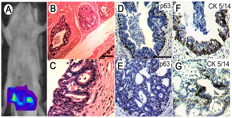
Figure 6. Orthotopic xenografting of PrCa/CCC cell cultures into the anterior prostate of recipient SCID mice recapitulated histological features of prostate adenocarcinoma.
(A) In vivo imaging of EGFP-labeled PrCa/CCC cells engrafted in the anterior prostates of recipient SCID mice shows localization of the grafts two weeks after grafting into the anterior prostate capsules. (B) Hematoxylin-eosin staining of tumor growth in prostate and urogenital organs. (C) Higher magnification of cribriform glands suggests initial development of invasive prostate cancer. Both simpler and more complex glands were observed. (D) Proliferation marker p63-stained basal cell layer of complex glands. (E) p63 expression appears disorderly in histologically higher grade glands. (F) Simpler glands express the marker CK5/14, whereas (G) glands that appear to be progressing to cribriform prostate cancer only sporadically express high molecular weight cytokeratin.

