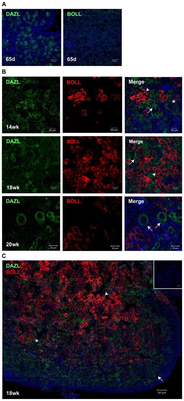
Figure 2. Dynamic changes of DAZL and BOLL protein expression during development of the human fetal ovary.
(A) at 65d gestation, DAZL is expressed in the nuclei and cytoplasm of germ cells in the human fetal ovary, but BOLL is not detected (B) At 14, 18 and 20 weeks gestation, DAZL is detected only in germ cell cytoplasm (arrows). BOLL-positive germ cells (arrowheads) are detectable from 14 weeks onwards, and increase in abundance with increasing gestation. Rare double-positive cells are marked by the asterisk. Primordial follicles at 20 weeks gestation (arrows, bottom left panel) express only DAZL. C) Tiled image of 18 weeks gestation fetal ovary section showing minimal co-localisation of DAZL and BOLL. DAZL is expressed in less mature germ cells in a more peripheral localisation, while BOLL predominantly expressed in more mature, centrally-located germ cells. Scale bar (A) and (B) 20 µm, (C) 50µm.

