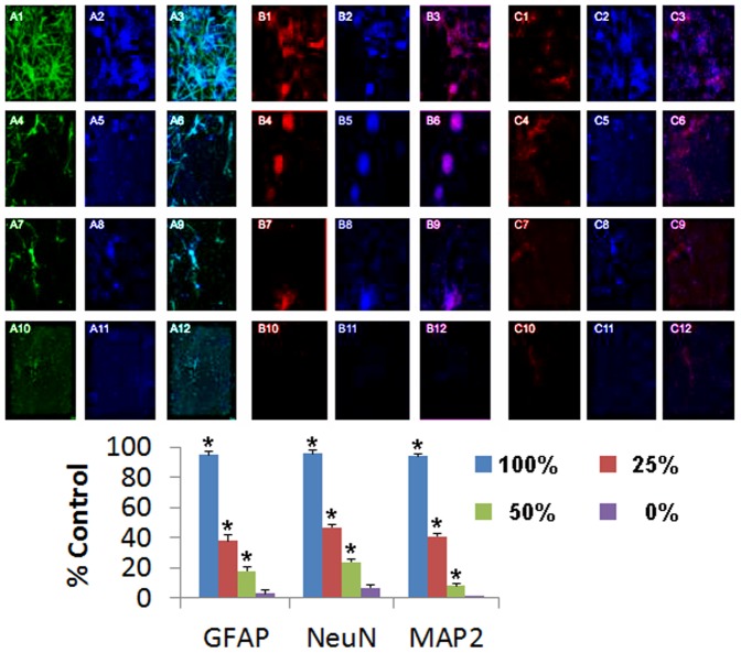
Figure 4. DPCs dose-dependently facilitate GFAP, NeuN, and MAP2 expression in primary rat cells post-OGD.
Immunocytochemical analyses revealed that co-culture of DPCs modulated dose-dependent expression of GFAP, NeuN, and MAP2 in OGD-exposed primary neural cells in the following order: 100%>50%>25%>0%. Phenotypic markers labeled as A: GFAP; B: NeuN; C: MAP2; Immunofluorescence identified as 1: phenotypic marker; 2: DAPI; 3: Merged; Cell doses given as 1–3: 100%; 4–6: 50%; 7–9: 25%; 10–12: 0%. Quantitative analyses revealed dose-dependent expression of these neural markers (*p<0.05 vs. controls).

