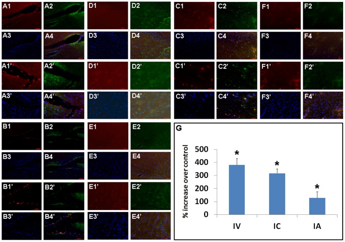
Figure 6. DPC grafts within the peri-infarct area co-localize with Hsp27 expression.
The majority of grafted DPCs were deposited within the ipsilateral peri-infarct striatal site as detected by the fluorescent dye tracker PKH26. Less than 1% of the grafts survived with no detectable differences in graft persistence whether delivered IV, IC, or IA (p>0.05). Only sparse PKH26-labeled cells were found in the contralateral intact hemisphere across all three delivery routes. Significantly increased Hsp27 expression was juxtaposed to grafted DPCs and quantitative analyses of Hsp27 expression (G) in the peri-infarct striatal site revealed robust Hsp27 expression in transplanted brains compared to controls, more pronounced in IV- and IC-delivered DPCs compared to IA-administered DPCs (*p<0.05 vs. controls). A: IV-delivered cells, ipsilateral; B: IC-delivered cells, ipsilateral; C: IA-delivered cells, ipsilateral; D: IV-delivered cells, contralateral; E: IC-delivered cells, contralateral; F: IA-delivered cells, contralateral; 1: PKH26; 2: Hsp27; 3: DAPI; 4: Merged. 1–4: 10X; 1′–4′:20×.

