
Figure 5. Role of AKT in the induction by TSH of IL-6.
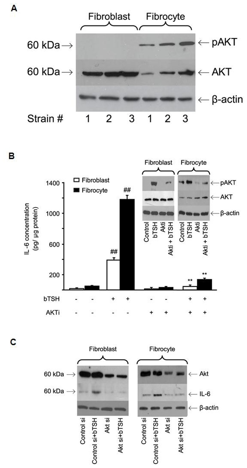
(A) Cellular proteins harvested from untreated cultures from 3 different donors each were subjected to Western blot analysis for AKT and pAKT Ser 473 as described in Experimental Procedures. (B) Confluent orbital fibroblast cultures, in this case from a patient with TAO, and fibrocytes from a healthy donor, were treated without or with bTSH (5 mIU/mL) in the absence or presence of AKTi (1 µM) for 16 hrs. Media were analyzed for IL-6 and cell layers for protein content. Data are presented as the mean ± SD of three independent determinations. (##, P<0.01 compared to untreated cultures; **, P<0.01 compared to TSH-treated cultures). In 3 separate experiments, Akti inhibited TSH-dependent IL-6 expression by 83±5% and 84±4% in fibroblasts and fibrocytes respectively. (Inset) TSH provokes AKT Ser 473 phosphorylation that is inhibited by AKTi. Cultures were treated with nothing or the agents indicated for 30 min. and cell protein was subjected to Western blotting. Densitometric analysis for pAKT bands: bTSH-treated fibroblasts, 29±3 AU; bTSH+AKTi, 9±2 AU. Fibrocytes, 34±5.5 AU and 14±2.7 AU, respectively. In 3 separate experiments, Akti inhibited pAKT by 67±7% and 58±9% in fibroblasts and fibrocytes, respectively. (C) knockdown of AKT with specific siRNA attenuates bTSH-induced IL-6. siRNA targeting AKT or control (scrambled) siRNA was transfected into 80% confluent cultures. After 48 h, these were treated without or with bTSH (5 mIU/mL) for 16 h. Protein was subjected to Western blot analysis. In 3 separate experiments, AKT siRNA reduced TSH-dependent IL-6 by 53±4% and 49±3% in fibroblasts and fibrocytes, respectively.