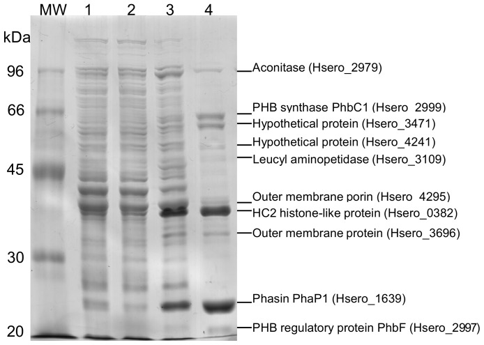
Figure 1. Electrophoretic pattern of PHB granule-associated proteins from Herbaspirillum seropedicae strain SmR1 in 10% SDS-PAGE.
Lanes: MW – molecular weight markers, 1– crude protein extract, 2– soluble fraction, 3– insoluble fraction and 4– granule-associated proteins after granule purification. Proteins were Coomassie blue R-250 stained.

