Figure 1.

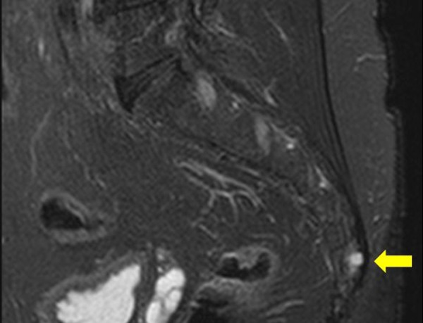
The sagittal T2 weighted MRI image of sacrococcygeal region, shows a small high signal lesion in the previous surgical site in the lower coccyx (arrow) consistent with recurrent chordoma.

The sagittal T2 weighted MRI image of sacrococcygeal region, shows a small high signal lesion in the previous surgical site in the lower coccyx (arrow) consistent with recurrent chordoma.