
Figure 4.
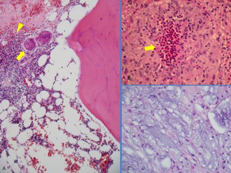
Photomicrographs of H&E staining images show: Astroid cells (arrow), and epithelioid granuloma (arrowhead) in the bone marrow (A), tumoral cells in the resected cocygeal bone (C), and epithelioid granuloma (arrow)in the resected lymph node (B).