Figure 1.
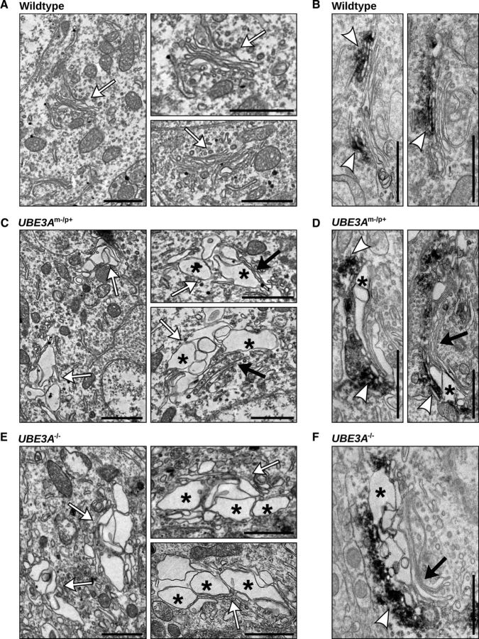
Disrupted morphology of the GA in UBE3Am−/p+ and UBE3A−/− mouse cortex. A–F, Electron micrographs of WT (A, B), UBE3Am−/p+ (C, D), and UBE3A−/− (E, F) neurons in primary visual cortex. The GA (white arrows) of WT neurons has tightly stacked cisternae with narrow intralumenal spaces arranged in stacked arrays. In contrast, the GA in UBE3Am−/p+ and UBE3A−/− mouse cortex contain enlarged and distended cisternae (asterisks), often adjacent to cisternae with normal morphology (black arrows). B, D, F, GM130 immunoreactivity (electron-dense DAB precipitates, white arrowheads) marking the cis-Golgi in WT (B), UBE3Am−/p+ (D), and UBE3A−/− (F) neurons of visual cortex. Scale bars, 1 μm.

