Figure 4.
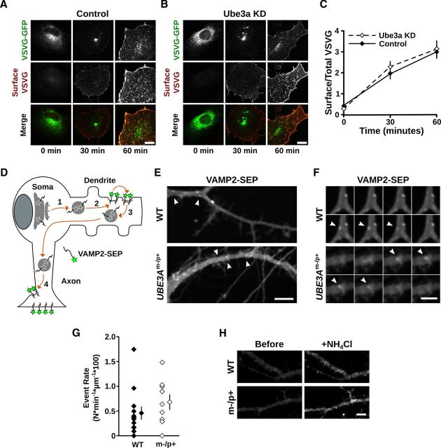
Secretory trafficking of VSVGts-GFP and VAMP2-SEP is not disrupted by loss of Ube3a. A–C, VSVGts-GFP progression from the ER to the plasma membrane through the GA was monitored after temperature-induced ER release (see Materials and Methods). VSVGts-GFP distribution and immunoreactivity at the plasma membrane (Surface VSVG) in (A) control and (B) Ube3a KD cells at 0, 30, and 60 min after release from ER-exit blockade. Scale bars, 10 μm. C, Quantification of surface VSVGts-GFP, measured by surface antibody staining, relative to total VSVGts-GFP, measured by intrinsic GFP fluorescence. No significant differences were evident in VSVG surface accumulation between control cells and Ube3a KD cells. Scale bars, 10 μm. p > 0.2 at all time points. D–H, Cargo transport from the GA to the plasma membrane was measured in UBE3Am−/p+ and WT cortical neurons by monitoring VAMP2-SEP exocytic events in dendrites. D, Schematic of VAMP2-SEP trafficking. Upon leaving the GA, newly synthesized VAMP2-SEP is trafficked into dendrites (1), where it is exocytosed at the plasma membrane (2). VAMP2-SEP is then rapidly endocytosed (3) and retrafficked to the axon, where it accumulates at axonal terminals (4). VAMP2-SEP fluoresces at neutral pH levels and is quenched at the acidic pH levels of intracellular organelles. E, Time-lapse projections (6.5 min) of WT and UBE3Am−/p+ neuronal dendrites expressing VAMP2-SEP. Arrowheads indicate exocytic events that occurred during the time-lapse. Scale bar, 10 μm. F, Time-lapse montage of selected exocytic events (arrowheads) from dendrites in E, one image per 5 s, showing the appearance and disappearance of VAMP2-SEP on the dendritic surface over time. Scale bar, 5 μm. G, Quantification of exocytic event rate (Nevents × min−1 × μm−1 × 100) in WT and UBE3Am−/p+ neurons; p = 0.28. H, Demonstration of the pH-sensitive properties of VAMP2-SEP in WT and UBE3Am−/p+ dendrites. There is weak diffuse VAMP2-SEP fluorescence under basal conditions and the much brighter, punctate VAMP2-SEP fluorescence after NH4Cl neutralizes pH in intracellular compartments. Scale bar, 10 μm.

