
Figure 5.
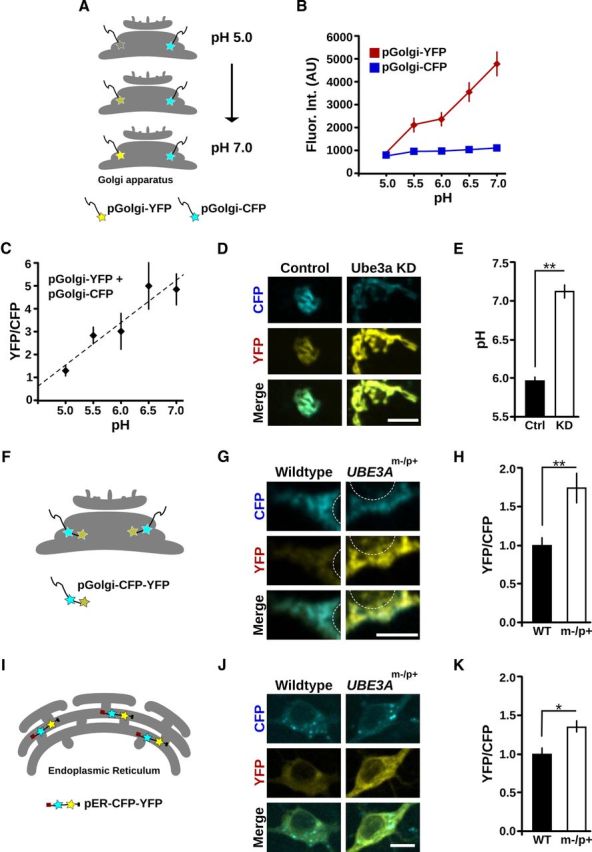
Golgi pH is elevated in Ube3a KD cells and in UBE3Am−/p+ neurons. A–E, Golgi pH is elevated in Ube3a KD cells. A, Schematic illustrating the method used to measure Golgi pH. YFP and CFP fluorophores are directed to the lumen of the GA using the Golgi targeting domain of β-1,4-galactosyltransferase. YFP fluorescence increases as pH increases, whereas CFP fluorescence is pH stable. B, C, Calibration of pGolgi-YFP:pGolgi-CFP ratios to absolute pH (see Materials and Methods). B, Quantification of pGolgi-YFP and pGolgi-CFP fluorescence (arbitrary units) at various defined pH values documenting their distinct pH sensitivities. C, Calibration curve (pGolgi-YFP:pGolgi-CFP fluorescence) used to calculate pH values. pH = 0.53 × YFP:CFP + 4.14, R2 = 0.9. D, pGolgi-YFP and pGolgi-CFP fluorescence in control and Ube3a KD cells. There is elevated pGolgi-YFP intensity in Ube3a KD cells compared with control cells. Scale bar, 10 μm. E, Quantification of Golgi pH in control (Ctrl) and Ube3a KD cells. **p < 0.001. F–H, Golgi pH is elevated in UBE3Am−/p+ neurons. F, Schematic of the probe used to measure Golgi pH in cultured cortical neurons. A Golgi pH probe with tandem CFP and YFP fluorophores (pGolgi-CFP-YFP) was expressed in cortical neurons cultured from WT and UBE3Am−/p+ mice. G, pGolgi-CFP-YFP fluorescence in WT and UBE3Am−/p+ neurons. White circles outline the nuclear region for orientation. There is elevated YFP intensity in the UBE3Am−/p+ neuron compared with the WT neuron. Scale bar, 10 μm. H, Average YFP:CFP fluorescence ratios of pGolgi-CFP-YFP in UBE3Am−/p+ neurons normalized to values in WT. **p = 0.001. I–K, ER pH is modestly elevated in UBE3Am−/p+ neurons. I, Schematic of the probe used to measure intralumenal ER pH. pER-CFP-YFP is a soluble protein targeted to the ER lumen by its N-terminal calreticulin signal sequence and C-terminal KDEL retrieval sequence. J, pER-CFP-YFP fluorescence in WT and UBE3Am−/p+ neurons. There is moderately elevated YFP intensity in the UBE3Am−/p+ neuron compared with the WT neuron. Scale bar, 10 μm. K, Average YFP:CFP fluorescence ratios of pER-CFP-YFP in UBE3Am−/p+ neurons after normalization to values in WT. *p < 0.01.