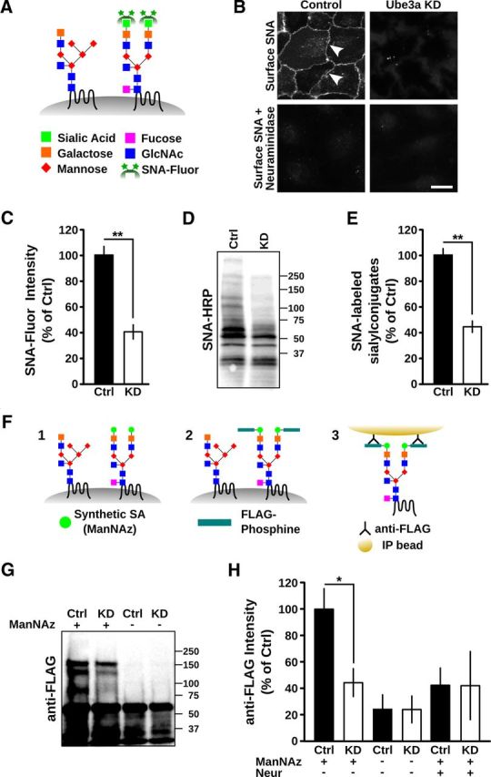
Figure 6.

Loss of surface protein sialylation in cells lacking Ube3a. A–E, Loss of α-2,6-sialic acid conjugates in Ube3a KD cells. A, Schematic illustrating labeling of α-2,6 linked sialic acid conjugates with SNA-fluorescein. GlcNAc, N-acetylglucosamine; Fluor, fluorescein. B, Surface labeling of α-2,6-sialic acid conjugates in control cells (cell membranes indicated with white arrowheads) and Ube3a KD cells using SNA-fluorescein. Pretreatment with neuraminidase abolished SNA labeling. Scale bar, 10 μm. C, Quantification of surface bound SNA-fluorescein on scrambled shRNA control (Ctrl) and Ube3a KD cells. **p < 0.001. D, Total cellular lysates from control and Ube3a KD cells after SDS-PAGE and blotting with SNA-HRP. Molecular mass markers are shown. E, Corresponding average integrated SNA-HRP signal demonstrating reduced sialylation in Ube3a KD cells. **p < 0.001. F–H, Reduction in total protein sialylation in Ube3a KD cells revealed by metabolic labeling. F, Schematic illustrating metabolic labeling and isolation of sialylated proteins using covalent FLAG-phosphine chemistry (see Materials and Methods). ManNAz, a synthetic azide derivative precursor to sialic acid (SA), is incorporated into endogenous glycans (F1), allowing reaction and covalent tagging with FLAG-phosphine (F2), and immunoprecipitation with an anti-FLAG antibody (F3). G, FLAG immunoreactivity after SDS-PAGE and immunoblotting of immunoprecipitates from control (Ctrl) and Ube3a KD cells with (+) or without (−) exposure to ManNAz. Molecular mass markers are shown. There are reduced levels of high molecular mass sialylconjugates in Ube3a KD cells and an absence of corresponding molecular species without prior metabolic labeling. The dark band at ∼55 kDa is the IgG band from the immunoprecipitation. Species <50 kDa are nonspecific as they are present in the absence of ManNAz. H, Average FLAG immunoreactivity (>60 kDa) in immunoblots, such as shown in G, and from samples exposed to ManNAz and treated with neuraminidase before FLAG-phosphine, defining background levels. There is reduction of protein sialylation in Ube3a KD cells. p > 0.3 for Ube3a KD versus negative controls (ANOVA). *p < 0.05 for Ube3a KD versus control.
