Figure 7.
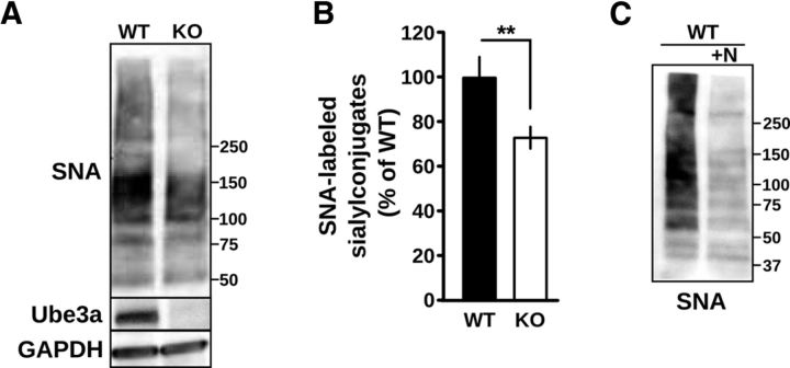
Reduction of α-2,6-sialylation in Ube3a-deficient cortex. A, Total cortical lysates from WT and UBE3A−/− mice after SDS-PAGE and blotting with SNA-HRP, anti-Ube3a, and anti-GAPDH. Molecular mass markers are shown. B, Corresponding average integrated signal of SNA-HRP reactivity in UBE3A−/− cortical lysates normalized to WT demonstrating a reduction in total α-2,6 sialylation. **p < 0.002. C, WT cortical lysates were incubated with neuraminidase (+N) to demonstrate the specificity of SNA-HRP for detection of sialylated residues. Molecular mass markers are shown.

