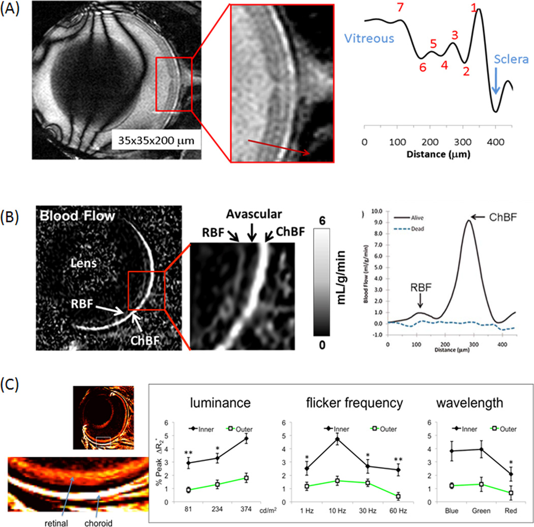
Figure 1.
(A) In vivo bSSFP MRI of the mouse retina at 45×45×500µm. Contrasts among different retinal layers are observed without exogenous contrast agents. Adapted from (11). (B) Layer-specific blood-flow image and blood-flow profile from a live mouse under 1.1% isoflurane and a dead mouse in the same setting (same animal) at 42×42×400µm. Blood flow was acquired using continuous arterial spin labeling with a separate labeling coil at the neck position. chBF – choroid blood flow, rBF – retinal blood flow. Adapted from (14). (C) Layer-specific blood-volume (MION) weighted images and fMRI responses to graded luminance, flicker frequency and wavelength (color) at 60×60×1000µm nominal resolution (mean ± SEM, N=7). Adapted from (22).

