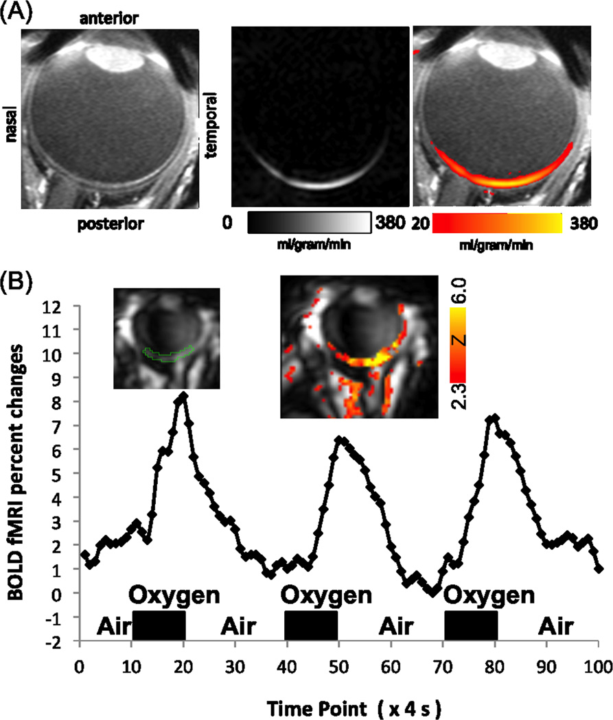
Figure 2.
(A) Blood-flow MRI of a human retina acquired using pseudo-continuous arterial spin labeling at 0.5×0.8×8mm (N=1). Adapted from (44). (B) BOLD fMRI map and time course of the human retina associated with oxygen versus air inhalation at 1.6×2×4mm (N=1). Inversion pulses were used to suppress the otherwise strong vitreous signal. Adapted from (46).

