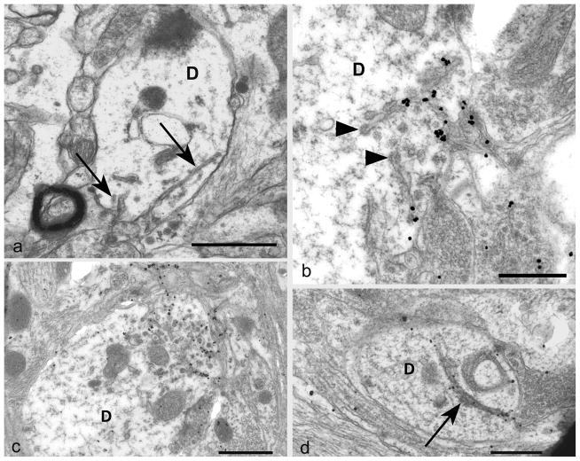
Fig 3. Plasma-lemmal membrane invaginations.
a) A dendrite (D) contains two plasma-lemmal membrane invaginations (arrows) the longer of which has a spiral arrangement. The dendrite lacks other sub-cellular organelles such as microtubules.
b) A tangentially sectioned dendrite (D) shows PrPSSLOW accumulation associated with coated tubular structures (arrowheads) and small membrane invaginations. PrPSSLOW is also present on membranes of adjacent processes.
c) A cross section of a dendrite (D) shows, at one pole, a marked increase in PrPSSLOW labelled coated pits and sub-plasma-lemmal fused tubular membrane complexes.
d) A large uncoated dendritic (D) membrane invagination (arrow) showing PrPSSLOW accumulation.
a: Uranyl acetate / lead citrate staining; b–d: immunogold labelling for PrPSSLOW
Mag bars: a = 1 μm, b = 0.5 μm, c = 1 μm, d = 0.5 μm.

