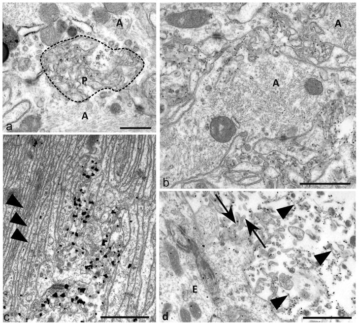
Fig 4. Plasma-lemmal membrane microfolding.
a) An early focus of plasma-membrane changes of astrocyte processes (A) showing ruffling of surface contours (bounded by dotted line) and irregular polyp like outfolds (P) of the plasma-membrane. Some cross or tangentially sectioned polyp-like folds have irregular ovoidal or elliptical profiles.
b) Prominent PrPSSLOW labelling of microfolded and polyp like extensions of astrocytic plasma-membranes. Larger astrocytic processes (A) from which these membrane changes arise are filled with intermediate filaments. PrPSSLOW is predominantly associated with membranes of microfolds.
c) An area of hyperplastic glial limitans of the pia showing parallel arrays of thin unlabelled astrocytic processes (arrowheads) with small areas of astrocytic microfolding showing PrPSSLOW accumulation.
d) PrPSSLOW labelling on microvilli (arrows) of ependymocytes (E) and on amyloid fibrils (arrowheads) within the ventricular lumen.
a: Uranyl acetate / lead citrate staining; b, c, d: immunogold labelling for PrPSSLOW Mag bars: a = 0.5 μm, b = 1 μm c = 1 μm, d = 1 μm.

