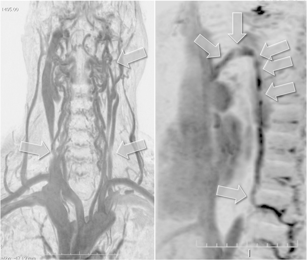
Figure 2.
Example of a Type A pattern as characterized by a stenosis or obstruction of the proximal azygous vein associated with a closed stenosis of one of the two internal jugular veins (IJVs), but with a compensatory contralateral IJV with apparent ample cross-sectional area. Both panels show subtraction masked, maximum intensity projection and inverted images from a 56 year old man with primary progressive multiple sclerosis that were obtained with dynamic contrast enhanced 3D fast field echo sequences. Arrows on the left hand panel point to a proximal left IJV stenosis and distal left and right IJV compression by sternocleidomastoid muscle that are not considered as hemodynamically significant. The right IJV is the dominant vessel with ample cross-sectional area. Arrows on the right hand panel point to multiple regions of narrowing of the azygous vein.

