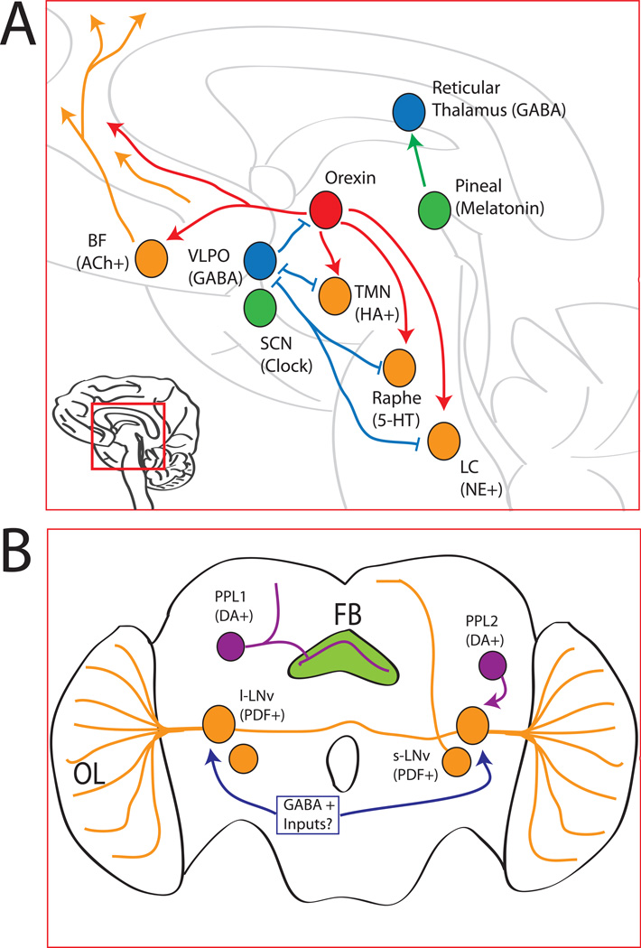
Figure 1. Major Sleep-Wake Pathways in Mammals and Flies.
A) The major mammalian sleep/wake regulatory pathways discussed in this review are shown. The ascending arousal system (ORANGE) is made up of many wake-promoting circuits, including the cholinergic basal forebrain (BF), the histaminergic (HA) tuberomammillary nucleus (TMN), the seroternergic (5-HT) dorsal raphe, and the noradrenaline (NE) producing locus coeruleus (LC). These areas send arousing projections (ORANGE LINES) to the thalamus and neocortex. The orexin neurons (RED) send excitatory projections to the ascending arousal network. The GABAergic neurons of the ventrolateral preoptic nucleus (VLPO; in BLUE), which makes inhibitory connections with both the orexin and ascending arousal systems. The TMN, raphe, and LC make mutual inhibitory connections with the VLPO to form a ‘flip-flop’ circuit. The pineal gland (GREEN) is the major source of melatonin, which can signal to the suprachiasmatic nucleus (SCN), which is the master regulator of circadian rhythms in mammals, and the GABA-positive reticular thalamus (BLUE), which may contribute to melatonin’s hypnotic effects. See text for details.
B) The major Drosophila circuits discussed in this review are shown on this schematic fly brain. Although not shown, all structures are bilaterally symmetric. In ORANGE are the major clock neurons, the large and small ventral lateral neurons (l-LNv and s-LNv). The wake promoting l-LNvs connect to the contralateral optic lobe (OL). Inhibitory GABA inputs (BLUE) are postulated for the l- LNvs. A single dopamine (DA) neuron (PURPLE) in the protocerebral posteriolateral 1 cluster (PPL1) signals to the fan shaped body (FB; GREEN) to increase Drosophila wakefulness. The wakepromoting l-LNvs also receive dopamine inputs, including from, but not limited to, the PPL2 cluster. See text for details.

