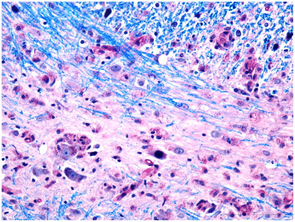
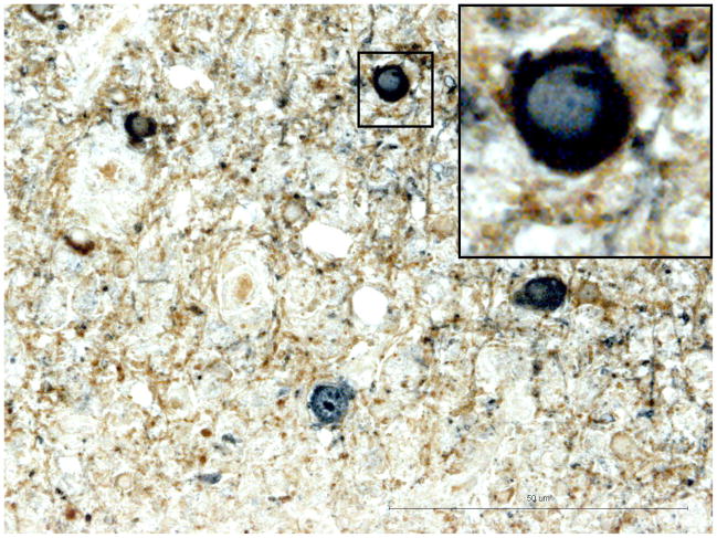
Fig. 2. Pons.
A) Intranuclear inclusion in oligodendroglial cells in a small focus of demyelination. Luxol Fast Blue combined with PAS. Additional inclusion at the edge of demyelination in upper part of the photograph. Bar 50 μm.
B) Double immunostaining for CNPase (Brown) and JCV Agno protein (blue). JCV antigen co-localizes with reaction for CNPase indicates infection of oligodendrocytes. Bar 50μ. Window – details of JCV infected oligodendroglial cell.


