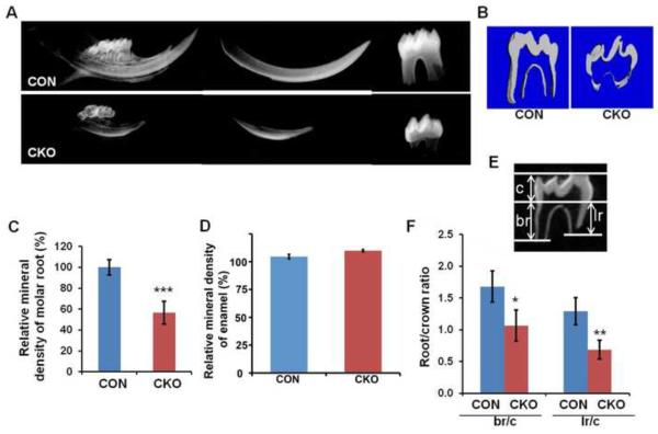
Figure 2. Root formation is disrupted in Tgfbr2CKO mice.
(A) Representative X-ray of mandibular incisors and the first molars from 3-week old control (CON) and Tgfbr2cko (CKO) mice. (B) Representative μCT images of the first mandibular molar from 3 weeks old CON and CKO mice. The parameters measured were (C) the mineral density in the root and (D) the mineral density of the enamel (E) Parameters used to evaluate tooth: crown (c), buccal root (br), and lingual root (lr) length. (F) The ratio of root length to crown length was calculated. Loss of Tgfbr2 in Osx-Cre expressing cells results in defects in root length, volume, and mineral density. Enamel density was not affected. N = 4; *: P < 0.05; **: P < 0.01; ***: P < 0.001.

