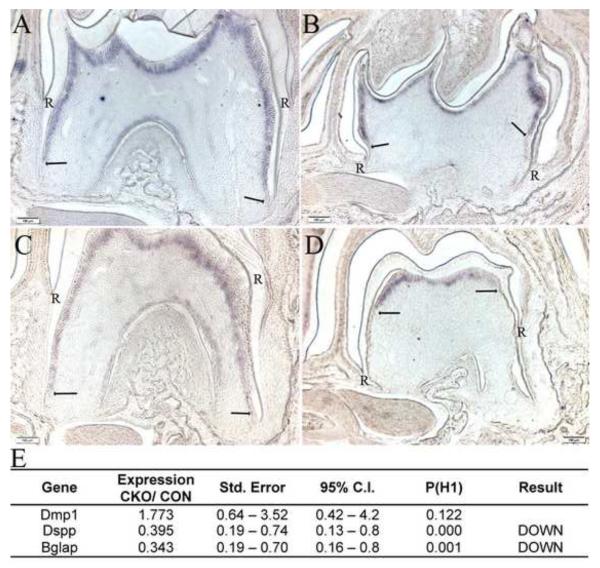
Figure 5. Odontoblast differentiation is disrupted in Tgfbr2CKO mice.
Representative images showing in situ hybridization for Dspp mRNA in the first mandibular molar form P10 (A, B) and P14 (C, D) day control (A, C) and Tgfbr2cko (B, D) mice. Hybridization to the Dspp probe is seen as purple staining. The termination of the expression domain is marked by arrows. Dspp expression observed in the lateral sides and bifurcation in control teeth is absent in the Tgfbr2cko teeth as is staining in the crown to root transition. R denotes where the root starts. (E) Expression of odontoblast markers in dental pulp of P10 day Tgfbr2cko mice (CKO) vs. control (CON) mice as determined by quantitative real time RT-PCR. Both beta-2-microglobulin (B2m) and glyceraldehyde 3 phospho dehydrogenase (Gapdh) were used as normalization controls.

