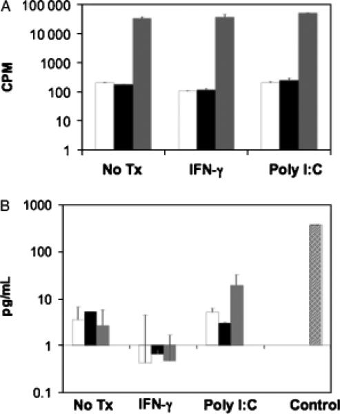
Fig. 4.
Functional antigen presentation of cultured cholangiocytes as measured by lymphoproliferation assay and interleukin (IL)-2 production. (A) Cholangiocytes functioned as experimental antigen-presenting cells (APCs) and were pretreated with media alone, poly I:C or interferon-γ (IFN-γ) before mitomycin C treatment. 2.5 × 10⁵ cholangiocytes were cultured with 2.5 × 10⁵ DO11.10 T cells in the absence (white bars) or presence (black bars) of 1 μg/ml ovalbumin (OVA)323–339 antigen. As a positive control, DO11.10 T cells were cultured with 2.5 × 10⁵ mitomycin C-treated bulk splenocytes with OVA antigen (grey bar). Cultures were pulsed with 1 μCi/well of thymidine-[methyl-3H] at 72 h and harvested 18 h thereafter. Values represent c.p.m. ± SEM of triplicate cultures. Pretreated cholangiocytes were unable to induce OVA antigen-specific T-cell proliferation compared with control APCs. (B) Cholangiocytes were pretreated with media alone, poly I:C or IFN-γ before mitomycin C treatment. 2.5 × 10⁵ cholangiocytes were cultured alone (white bar), or cocultured with 2.5 × 10⁵ OVA-specific DO11.10 hybridoma cells and without (black bars) or with OVA antigen (grey bars). As a positive control, DO11.10 hybridoma cells were cultured with 2.5 × 10⁵ mitomycin C-treated bulk splenocytes with OVA antigen (hatched bar). Culture supernatants were collected at 24 h and analysed by enzyme-linked immunosorbent assay for IL-2 protein. Values represent the mean pg/ml ± SEM. Cholangiocytes were unable to induce IL-2 secretion from DO11.10 hybridomas in the presence of OVA antigen compared with control APCs.

