Figure 3.

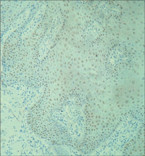
Moderate to strong immunostaining of areas of moderate dysplasia with cyclin D1 in basal, parabasal and suprabasal cells along with numerous mitotic figures in the spinous layer under ×10 magnification

Moderate to strong immunostaining of areas of moderate dysplasia with cyclin D1 in basal, parabasal and suprabasal cells along with numerous mitotic figures in the spinous layer under ×10 magnification