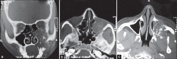
Figure 2.

(a) Coronal CT view depicting an expansile lesion of the right maxilla with expansion and thinning of the overlying buccal cortex and involvement of the right maxillary antrum. Erosion of the medial wall of the orbit was also seen (b) Axial CT view showing extension of the lesion into the nasal cavity (c) Axial CT view depicting irregular calcific strands within the lesion
