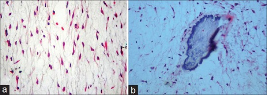
Figure 3.

(a) Photomicrograph of the histopathologic section reveals stellate shaped fibroblastic cells set in a myxoid background with delicate haphazardly arranged collagen fibers (H and E, ×200) (b) Photomicrograph of the histopathologic section reveals foci of residual bone within myxoid background (H and E, ×200)
