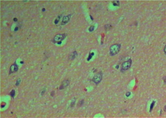
Fig. 1.

Micrograph of brain section of gonad intact control rat showing the highly active nerve cells that having huge nuclei with relatively pale-stained, the nuclear chromatin and prominent nuclei disappeared. The surrounding support cells having small nuclei with densely stained, condensed chromatin with no visible nucleoli are shown in the cortex (H and E ×400)
