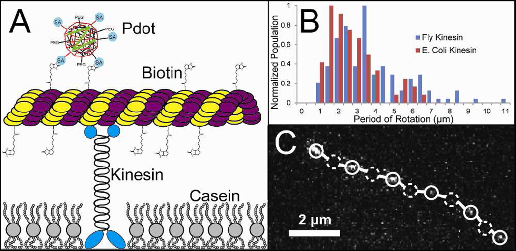
Figure 4.
Polarization sensitive polymer nanoparticles used to detect microtubule rotation. (A) scheme showing a gliding microtubule moved by kinesin bound to a glass substrate. The polymer nanoparticles link to biotinylated tubulin within the microtubule. (B) measured microtubule periods of rotation when the microtubules are transported by two different forms of kinesin. There were 62 microtubules measured using E. coli kinesin and 131 measured using Drosophila kinesin along with 64 and 148 non-rotating microtubules, respectively. (C) Track of a single microtubule with a single bound polymer nanoparticle (open circles) show local intensity maxima while dashed circles indicate the location of local intensity minima in one of the observed channels. The optical setup used to capture the image track is described in reference 19.

