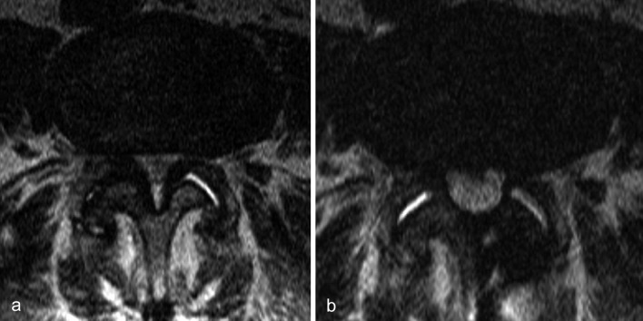
Figure 3.
Bilateral, severe spinal canal and lateral recess stenosis
a) Preoperative axial T2-weighted MRI at the L3 and L4 levels
b) An MRI six months after surgery reveals the extent of decompression, performed in this case as a unilateral interlaminar decompression from the left with undercutting on the right. The typical spinal claudication that was present before surgery resolved completely. Back pain on movement remained to some extent but did not cause any significant impairment. (With kind permission of the Neuroradiology Division at the Universitätsklinikum Jena)

