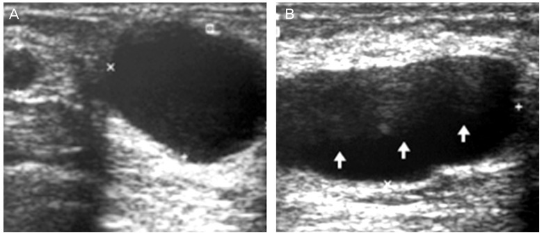
Fig. 10.

Galactocele 6 months post delivery which shows various features. (A) Ultrasound (US) image reveals 2 cystic mass: typical galactocele with homogenous anechoic, acoustic attenuation and lateral edge shadowing in bigger cyst. (B) US image shows the lobulated, fat-fluid (arrows) galactocele.
