Fig. 1.

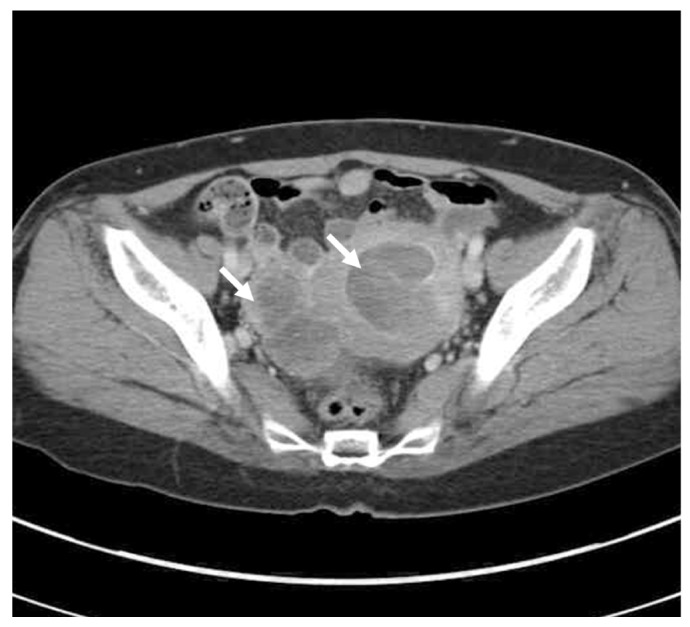
Computed tomography showing a huge sized mass in uterine cavity. And 5.5-cm sized cystic and solid mass in right adnexa (white arrows).

Computed tomography showing a huge sized mass in uterine cavity. And 5.5-cm sized cystic and solid mass in right adnexa (white arrows).