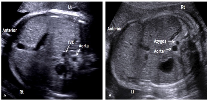
Fig. 4.
Transverse abdominal views of both forms of heterotaxy syndrome. (A) Fetus with right atrial isomerism (breech presentation) shows juxtaposition of the aorta and IVC on either left side of the spine. The right-sided stomach and the midline liver are also shown. (B) Fetus with left atrial isomerism (vertex presentation) shows invisible IVC with dilated azygos vein, suggestive of an interrupted IVC with azygos vein continuation. Lt, left; Rt, right; IVC, inferior vena cava.

