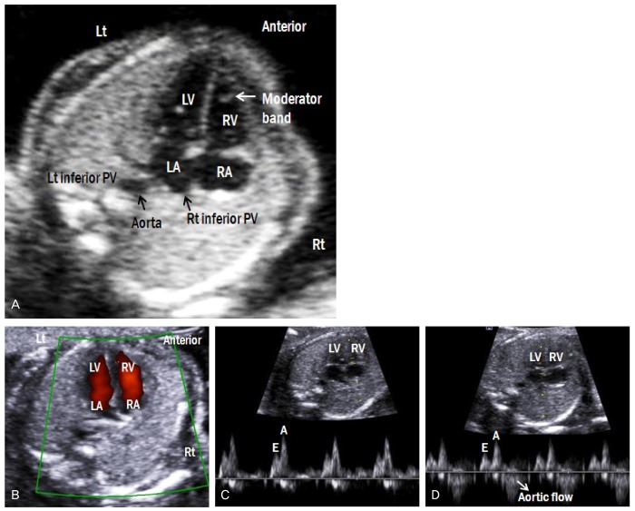
Fig. 5.
(A) Apical four-chamber view of the fetal heart. (B) Color Doppler image shows forward flow from both atria to both ventricles. (C) Doppler waveforms of tricuspid and (D) mitral valves show a biphasic pattern. The E wave corresponds to early ventricular filing of the diastole, and the A wave corresponds to active ventricular filling of the diastole (atrial contraction). (D) Because of the continuity between mitral and aortic valves, the Doppler waveform at the mitral valve shows aortic outflow. Lt, left; Rt, right; LA, left atrium; RA, right A atrium; LV, left ventricle; RV, right ventricle; PV, pulmonary vein.

