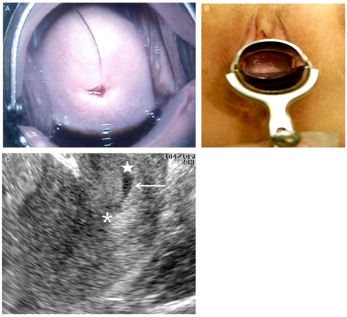
Fig. 1.
(A) Before allogenic peripheral blood stem cell transplantation, photograph shows normal uterine cervix with intra-uterine device tail string which got stuck into exocervix. (B) Twenty-six months after allogenic peripheral blood stem cell transplantation, photograph shows almost complete obstruction of the upper vaginal canal. A lower vagina had normal appearance. (C) Twenty-six months after allogenic peripheral blood stem cell transplantation, trans-rectal ultrasonography shows hematocolpos (arrow). asterisk, uterine cervix; star, vagina.

