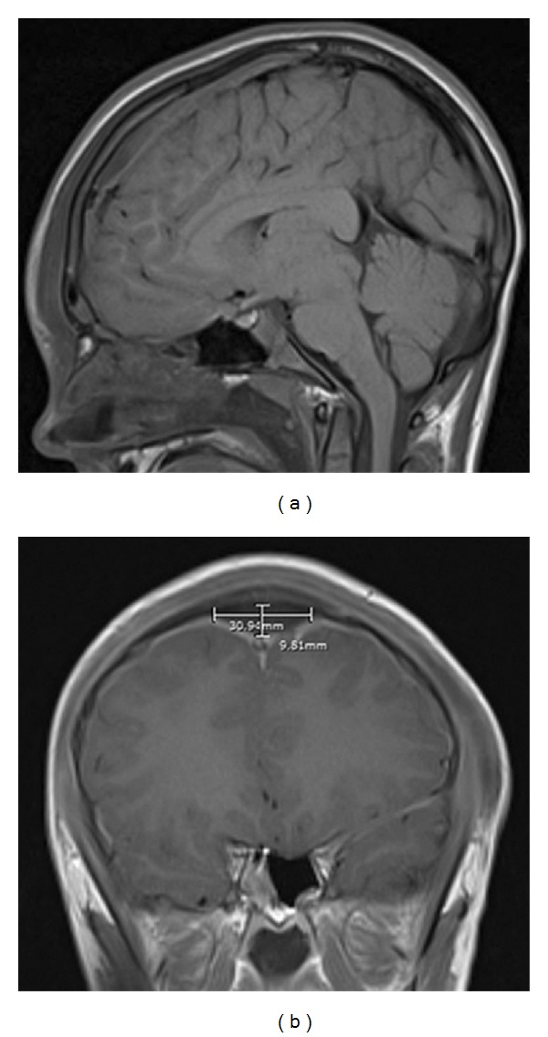
Figure 1.

(a) MRI of the brain showing sagittal T1-weighted image after administration of contrast. The fluid collection reported to have a thin enhancing wall ran along the dorsal aspect of the superior sagittal sinus. (b) MRI of the brain showing coronal T1-weighted image after administration of contrast. Diffuse smooth dural enhancement is noted, bilaterally.
