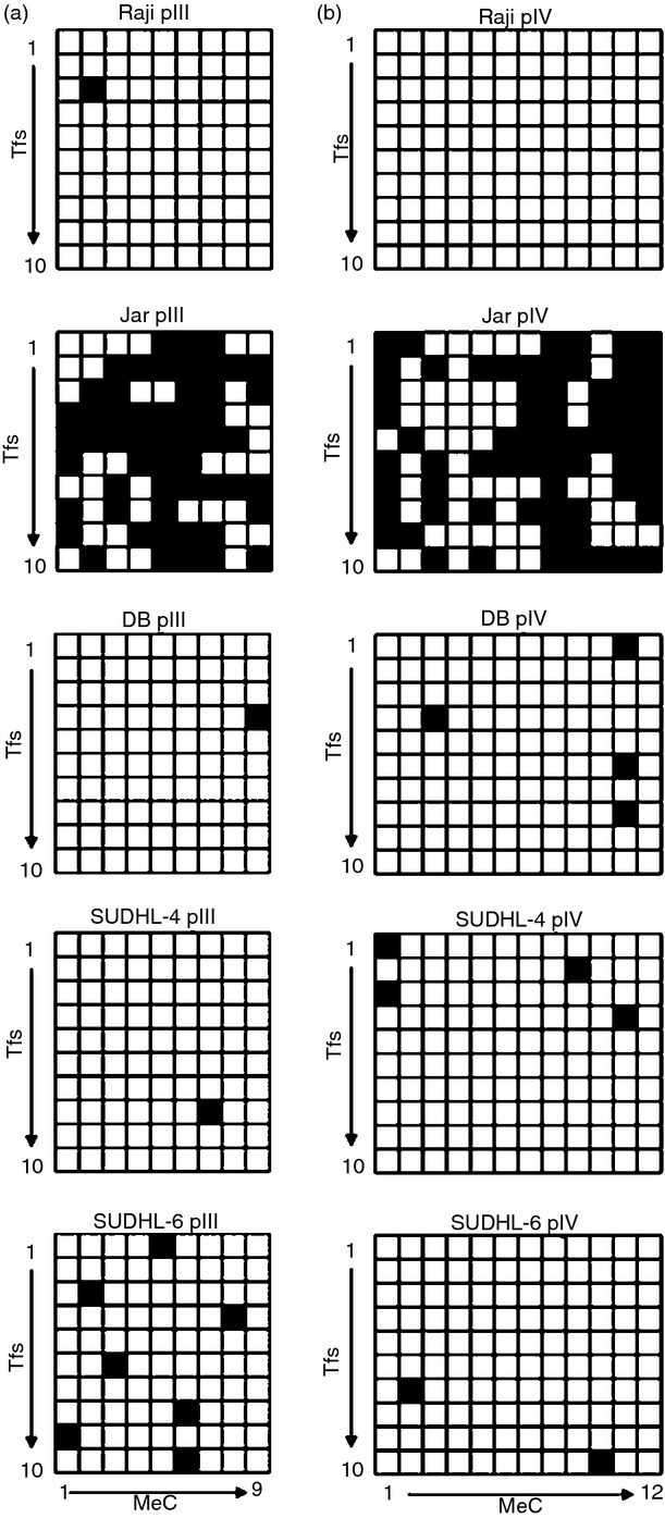
Figure 3.

DNA methylation analysis at CIITA promoters III and IV (pIII, pIV) in diffuse large B-cell lymphoma (DLBCL) cells. Genomic DNA was isolated from DB, SUDHL-4, and SUDHL-6 cells and subjected to bisulphite sequencing analysis for methylated CpG dinucleotides (MeC) within (a) CIITA pIII (455 bp region) and (b) pIV (362 bp region). These CIITA pIII and pIV regions contain 9 and 12 CpG dinucleotides, respectively (x-axis). Ten transformants (y-axis) were sequenced for each CIITA promoter in each cell line. Black and white squares represent methylated and unmethylated CpG dinucleotides, respectively. Raji and Jar cells (shown) and Jurkat cells were included as negative and positive controls, respectively, for CpG methylation at the CIITA promoters.
