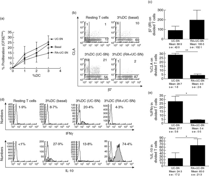
Fig. 6.
Dendritic cells (DC) conditioned with retinoic acid (RA) maintain their homeostatic, gut-specific properties in the presence of a proinflammatory microenvironment. (a) Monocyte-derived DC were conditioned with culture supernatants from inflamed intestinal biopsies of ulcerative colitis patients [(UC)-supernatants (SN)] with and without previous conditioning with RA and compared to untreated (basal) DC. T cell proliferation following mixed lymphocyte reaction (MLR) of carboxyfluorescein diacetate succinimidyl ester (CFSE)-labelled T cells with allogeneic DC was determined as explained in Fig. 4a. (b) Gut-homing (β7) and skin-homing [cutaneous lymphocyte-associated antigen (CLA)] markers were assessed on resting T cells (unstimulated) and on T cells (CFSElow) stimulated with DC preconditioned with medium only, UC-SN or RA and UC-SN. (c) Pooled data of tissue-homing profile of stimulated T cells. (d) Intracellular production of interleukin (IL)-10 and interferon (IFN)-γ by stimulated T cells following MLR and (e) pooled data of cytokine production by stimulated T cells. Graphs show the mean ± standard error of the mean of three independent experiments. Two-way anova (a) and paired t-test (c,e) were applied. P-value below 0·05 was considered statistically significant. *P < 0·05; **P < 0·01.

