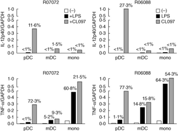
Fig. 8.

Interleukin (IL)-12p40 and tumour necrosis factor (TNF)-α mRNA expression in Toll-like receptor (TLR)-4 (lipopolysaccharide) and TLR-7/8 (CL097) stimulated purified rhesus macaque plasmacytoid dendritic cells (pDC), myeloid dendritic cells (mDC) and monocytes. Shown are expression levels of IL-12p40 (top graphs) and TNF-α (bottom graphs) relative to glyceraldehyde 3-phosphate dehydrogenase (GAPDH) in pDC, mDC and monocytes as well as percentage of positive cells as detected by fluorescence activated cell sorter (FACS) analysis on the same cells (number at top of bars). Results from two individual rhesus macaques are shown.
